The synthesis of medical image acquisition and principles of image contrast formation can yield powerful diagnostic tools. A classic example in MRI is automated fat-water separation, and myriad related strategies to quantify tissue fat content. A historical method is illustrated, where phase differences observed on dual-echo, gradient echo images are disambiguated by a region growing algorithm–into mostly fat, or mostly water subregions.
3D Isotropic fast spin echo imaging
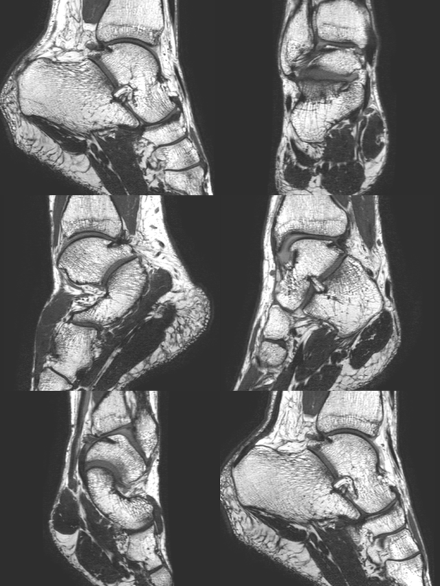
We first promoted this idea over a decade ago, and it has caught on in only limited applications. While techniques need to be tailored to clinical applications. isotropic TSE is well suited as a comprehensive, high resolution evaluation of small parts in musculoskeletal imaging, where it may be the one application that leverages the full benefits of expensive, multi-channel coil technology.
Quantitative Imaging
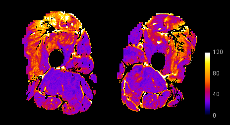
Expert image interpretation is predicated on knowledge and experience. The use of imaging as a biomarker in medical research could be empowered by leveraging methods and concepts that remove the subjectivity of image interpretation. For example, we have pioneered the use of muscle T2 maps, corrected for fat content, as a marker of active disease in inflammatory myopathy.


